Free Online PDB Viewer (No Signup)
Also opens DWG, DXF, STEP, IGES, IFC, SKP, STL, OBJ, GLB, FBX, 3DM and other 3D/CAD files. Drop any file on the viewer.
Works directly in Chrome, Firefox, Safari, and Edge
Also from Innerscene: Circadian Lab, a free lighting calculator for room illuminance, melanopic EML, and glare.
Describe a compound, a protein or a crystal. The AI finds it in PubChem or the Protein Data Bank, or builds it, and opens it here.
Create a model with AI
Describe what you need, or add a photo, and AI builds it at real size in the viewer. Ask for changes in the chat, then export it. Free, with your own Anthropic or OpenAI key.
Open the AI GeneratorWhat Is a PDB File?
A .pdb file is plain text in fixed columns. ATOM and HETATM records give each atom its name, element, residue, chain and coordinates; HELIX and SHEET records mark the secondary structure; CONECT records list bonds for ligands and unusual residues; CRYST1 gives the crystal’s unit cell; and MODEL blocks hold the models of an NMR ensemble. The format holds up to 99,999 atoms and 62 chains, so the largest entries in the Protein Data Bank are published in PDBx/mmCIF only.
Three variants share the layout. .ent is the name the wwPDB archive gives its PDB files (pdb1abc.ent). PDBQT, written by AutoDock Tools, Open Babel and Meeko for AutoDock and AutoDock Vina, adds partial charges and AutoDock atom types for docking receptors and ligands. PQR, written by PDB2PQR for APBS, puts each atom’s charge and radius in the columns PDB uses for occupancy and temperature factor.
For small molecules, see the SDF and MOL viewer; for every molecular format on one page, the molecule viewer.
Who Uses PDB Files?
Structural biologists and biochemists, drug-discovery teams reading docking results, students and teachers in biology and chemistry classes, bioinformaticians, science illustrators, and designers who want a real protein for a render, an animation or a 3D-printed model.
Common Use Cases
- Look at a protein from the RCSB PDB, PDBe or PDBj without installing PyMOL or ChimeraX
- Check a docking result: the receptor and the docked ligand from AutoDock Vina as PDBQT
- Show a structure to a class or a client as a link that opens in any browser
- Export a protein as STL or 3MF for a 3D-printed model
- Bring a protein into Blender, a game engine or an AR scene as GLB, FBX or USDZ
Features
Opens .pdb Files
Protein Data Bank structures, wwPDB .ent files, AutoDock PDBQT and APBS PQR.
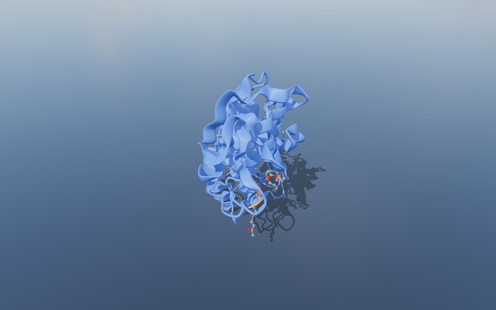
Cartoon of Every Chain
Helices and strands as ribbons from the file, or worked out from the Cα trace; one chain coloured blue to red, several chains each in its own colour.
DNA and RNA
Nucleic acids as a tube along the backbone with the bases as sticks.
Ligands as Ball and Stick
Bound ligands, cofactors and ions drawn as ball and stick, coloured by element.
Layers to Switch On
Polymer atoms as sticks, hydrogens, water, space filling and the unit cell, each on its own layer.
Export to GLB, STL or USDZ
Save the structure as GLB, glTF, OBJ, STL, 3MF, PLY, FBX or USDZ for a render, a 3D print or AR, or the view as a PNG.
Measure in Ångströms
Click two atoms to measure the distance between them, in Å.
View in VR
On a headset browser the molecule stands on a table in front of you.
Nothing Is Uploaded
The file is read entirely in your browser. No account, no upload, no copy on a server.
Share and Embed
Send a link that opens the molecule in any browser, or drop the viewer into your own page with one iframe.
Compatibility & Format Notes
ATOM, HETATM, HELIX, SHEET, CONECT and CRYST1 records are read. The element comes from its own column, or from the atom name when that column is blank (as in many older files and docking outputs). Only the first model of a multi-model file and the first alternate location of each atom are drawn. PDBQT and PQR files open the same way, with their extra columns read past.
Proteins open as a cartoon: helices and strands as ribbons, taken from the file’s secondary-structure records when it has them and worked out from the Cα trace when it does not, with loops as a round tube. A single chain is coloured along its sequence from blue at the start to red at the end; when there are several chains, each gets its own colour. DNA and RNA open as a tube along the backbone with the bases as sticks. Ligands are drawn as ball and stick, coloured by element (carbon grey, oxygen red, nitrogen blue, sulfur yellow, in the Jmol colours).
Each representation is a layer you can switch on and off: Cartoon, Polymer atoms (every heavy atom of the chains as sticks), Hydrogens, Ligands, Water, Space filling (every atom except water as a sphere at its van der Waals radius) and Unit cell when the file gives one. Cartoon and ligands are on when the file opens; the rest wait in the Layers panel.
Coordinates are read in ångströms (Å) and kept at that scale, so the measuring tool reads distances in Å. Bonds are taken from the file where it lists them and worked out from covalent radii for the rest. Very large structures, such as a whole virus capsid with millions of atoms, open with the cartoon and with their atoms drawn as points so the browser keeps up.
Embed on Your Website
Add this viewer to your own site with a single line of HTML. Click Embed in the toolbar to generate an embed code, or use the snippet below with your own file URL.
Your Files Stay on Your Computer
Your PDB files are parsed entirely in your browser. Nothing is uploaded, stored, or shared unless you explicitly click the Share button. Shared files are hosted for 90 days then deleted.
Frequently Asked Questions
▶How do I open a structure from the Protein Data Bank?
On the entry’s page at rcsb.org, PDBe or PDBj, choose Download Files and pick PDB Format (or PDBx/mmCIF for large entries), then drop the file on the viewer. Both formats open.
▶Why is my protein shown as ribbons?
The cartoon shows the fold of each chain at a glance. To see every atom, switch on Polymer atoms (sticks) or Space filling (spheres) in the Layers panel, and switch off Cartoon if you like.
▶Do PDBQT and PQR files open?
Yes. AutoDock and AutoDock Vina PDBQT files (receptors, ligands and docked poses) and APBS PQR files open like PDB files. A PDBQT file with several docked poses shows the first one.
▶Which model of an NMR structure is shown?
The first model of the ensemble. The other models are read past.
▶Where are the hydrogens and the water?
They are on their own layers, Hydrogens and Water, which start switched off so the structure stays readable. Switch them on in the Layers panel.
▶Can I 3D print the molecule?
Yes. Export STL or 3MF and open it in your slicer. Thin cartoon ribbons and sticks are fragile at small sizes, so for a sturdy print switch on Space filling (or ball and stick for a small molecule) before exporting, and scale the model up in the slicer: one ångström in the file is one unit in the export.
▶Is this PDB viewer really free?
Yes, completely free. No signup, no watermarks, no file size limits on the viewer (the optional share-link feature has a 100 MB upload cap). Built by Innerscene as a tool for architects, engineers, and designers.
▶Are my files uploaded to a server?
No. All parsing, rendering, and conversion runs in your browser: nothing is uploaded, stored, or transmitted by default. The Share button is the only feature that uploads a file, and only when you explicitly click it.
▶Can I convert to another format?
Yes. Click Export in the toolbar to save the structure as GLB, glTF, OBJ, STL, 3MF, PLY, FBX or USDZ, or the view as a PNG picture. The export carries the layers you have switched on, so turn on Space filling or Polymer atoms first if you want them in the file. There is no PDB or SDF writer: the exports are 3D models of the picture you see.
▶Can I embed this viewer on my own site?
Yes. Click Embed in the toolbar to generate a one-line <iframe> snippet you can paste into any HTML page. You can choose whether the embed shows the viewer’s toolbar.