Free Online CIF Viewer (No Signup)
Also opens DWG, DXF, STEP, IGES, IFC, SKP, STL, OBJ, GLB, FBX, 3DM and other 3D/CAD files. Drop any file on the viewer.
Works directly in Chrome, Firefox, Safari, and Edge
Also from Innerscene: Circadian Lab, a free lighting calculator for room illuminance, melanopic EML, and glare.
Describe a compound, a protein or a crystal. The AI finds it in PubChem or the Protein Data Bank, or builds it, and opens it here.
Create a model with AI
Describe what you need, or add a photo, and AI builds it at real size in the viewer. Ask for changes in the chat, then export it. Free, with your own Anthropic or OpenAI key.
Open the AI GeneratorWhat Is a CIF File?
A CIF file is a list of named items and tables (loop_ blocks) in the STAR syntax. Two dictionaries use it. PDBx/mmCIF is the archive format of the Protein Data Bank: _atom_site lists every atom with its residue, chain and Cartesian coordinates, _struct_conf and _struct_sheet_range mark helices and strands, and _struct_conn lists links between residues and ligands. It has no limit on atoms or chains, so large complexes such as ribosomes are published in mmCIF only.
Small-molecule CIF is what crystallographers deposit with the Cambridge Crystallographic Data Centre (CCDC), the Crystallography Open Database (COD) and IUCr journals. It gives the unit cell (_cell_length_a and the angles), the space group and the fractional coordinates of the atoms of the asymmetric unit.
For the older Protein Data Bank format, see the PDB viewer; for every molecular format on one page, the molecule viewer.
Who Uses CIF Files?
Crystallographers and structural biologists, chemists and materials scientists reading structures from the CCDC or the COD, students in chemistry, mineralogy and biology, and designers and architects who want the geometry of a real crystal or protein for a render, a pattern or a 3D print.
Common Use Cases
- Open a large Protein Data Bank entry that is only published as PDBx/mmCIF
- Look at a crystal structure from the COD or a journal’s supporting information with its unit cell
- Check a structure before a group meeting or a lecture without opening Mercury, PyMOL or ChimeraX
- Share a crystal or protein as a link that opens in any browser
- Export the structure as STL, 3MF, GLB or OBJ for a print, a render or a game engine
Features
Opens .cif Files
PDBx/mmCIF from the Protein Data Bank and small-molecule CIF from the CCDC, the COD and journals.
Crystals in Their Unit Cell
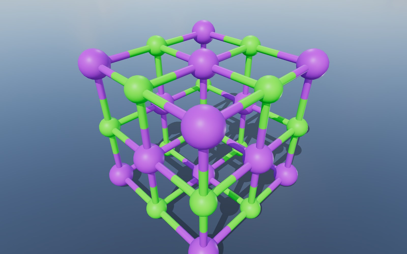
Fractional coordinates turned into ångströms, with the unit cell drawn as a box around the atoms.
Cartoon of Every Chain
Helices and strands as ribbons from the file, or worked out from the Cα trace; one chain coloured blue to red, several chains each in its own colour.
DNA and RNA
Nucleic acids as a tube along the backbone with the bases as sticks.
Ligands as Ball and Stick
Bound ligands, cofactors and ions drawn as ball and stick, coloured by element.
Layers to Switch On
Polymer atoms, hydrogens, water, space filling and the unit cell, each on its own layer.
Export to GLB, STL or USDZ
Save the structure as GLB, glTF, OBJ, STL, 3MF, PLY, FBX or USDZ for a render, a 3D print or AR, or the view as a PNG.
Measure in Ångströms
Click two atoms to measure the distance between them, in Å.
View in VR
On a headset browser the molecule stands on a table in front of you.
Nothing Is Uploaded
The file is read entirely in your browser. No account, no upload, no copy on a server.
Share and Embed
Send a link that opens the molecule in any browser, or drop the viewer into your own page with one iframe.
Compatibility & Format Notes
The first data block with atom coordinates is read (a leading block of publication details is skipped). In mmCIF, _atom_site (first model, one alternate location per residue), _struct_conf, _struct_sheet_range and _struct_conn are used. In a small-molecule CIF, the unit cell and the fractional coordinates are turned into Cartesian ångströms; the atoms drawn are those the file lists (the asymmetric unit), and the unit cell is drawn as a box around them.
Proteins open as a cartoon: helices and strands as ribbons, taken from the file’s secondary-structure records when it has them and worked out from the Cα trace when it does not, with loops as a round tube. A single chain is coloured along its sequence from blue at the start to red at the end; when there are several chains, each gets its own colour. DNA and RNA open as a tube along the backbone with the bases as sticks. Ligands are drawn as ball and stick, coloured by element (carbon grey, oxygen red, nitrogen blue, sulfur yellow, in the Jmol colours).
Each representation is a layer you can switch on and off: Cartoon, Polymer atoms (every heavy atom of the chains as sticks), Hydrogens, Ligands, Water, Space filling (every atom except water as a sphere at its van der Waals radius) and Unit cell when the file gives one. Cartoon and ligands are on when the file opens; the rest wait in the Layers panel.
A small-molecule crystal opens as ball and stick with its Unit cell layer switched on; for a macromolecule the unit cell starts hidden.
Coordinates are read in ångströms (Å) and kept at that scale, so the measuring tool reads distances in Å. Bonds are taken from the file where it lists them and worked out from covalent radii for the rest. Very large structures, such as a whole virus capsid with millions of atoms, open with the cartoon and with their atoms drawn as points so the browser keeps up.
Embed on Your Website
Add this viewer to your own site with a single line of HTML. Click Embed in the toolbar to generate an embed code, or use the snippet below with your own file URL.
Your Files Stay on Your Computer
Your CIF files are parsed entirely in your browser. Nothing is uploaded, stored, or shared unless you explicitly click the Share button. Shared files are hosted for 90 days then deleted.
Frequently Asked Questions
▶Does it open both mmCIF and small-molecule CIF?
Yes. A PDBx/mmCIF file from the Protein Data Bank opens as a cartoon of its chains with ligands; a crystallography CIF from the CCDC, the COD or a journal opens as ball and stick inside its unit cell. The viewer tells them apart from the content.
▶Is the crystal packing generated from the symmetry?
No. The atoms drawn are the ones the file lists, which for a small-molecule CIF is the asymmetric unit, with the unit cell drawn around them.
▶My CIF file holds several structures. Which one opens?
The first one with atom coordinates; a block of publication details at the top is skipped. To see another structure, save its block as its own file.
▶Can I open the very large structures that have no PDB file?
Yes. Ribosomes, viral capsids and other entries published only as mmCIF open with their cartoon; past a quarter of a million atoms in a layer, the atoms are drawn as points so the browser keeps up.
▶Where are the hydrogens and the water?
On their own layers, Hydrogens and Water, which start switched off. Switch them on in the Layers panel.
▶Can I 3D print the molecule?
Yes. Export STL or 3MF and open it in your slicer. Thin cartoon ribbons and sticks are fragile at small sizes, so for a sturdy print switch on Space filling (or ball and stick for a small molecule) before exporting, and scale the model up in the slicer: one ångström in the file is one unit in the export.
▶Is this CIF viewer really free?
Yes, completely free. No signup, no watermarks, no file size limits on the viewer (the optional share-link feature has a 100 MB upload cap). Built by Innerscene as a tool for architects, engineers, and designers.
▶Are my files uploaded to a server?
No. All parsing, rendering, and conversion runs in your browser: nothing is uploaded, stored, or transmitted by default. The Share button is the only feature that uploads a file, and only when you explicitly click it.
▶Can I convert to another format?
Yes. Click Export in the toolbar to save the structure as GLB, glTF, OBJ, STL, 3MF, PLY, FBX or USDZ, or the view as a PNG picture. The export carries the layers you have switched on, so turn on Space filling or Polymer atoms first if you want them in the file. There is no PDB or SDF writer: the exports are 3D models of the picture you see.
▶Can I embed this viewer on my own site?
Yes. Click Embed in the toolbar to generate a one-line <iframe> snippet you can paste into any HTML page. You can choose whether the embed shows the viewer’s toolbar.